Biomolecules:
Protein 1
Planarity of Peptide Bonds
Interestingly, peptide bonds have a second resonance form, as demonstrated below. This means that the peptide bond (the C=O and N-H) all reside in a single plane. Thus, there is no rotation around the
|
|
|
|
|
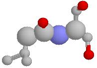
The structure at the right shows a peptide bond between the amino acids valine (Val) and serine (Ser). The amino acids are taken from the crystal structure of hemoglobin (αVal132 and αSer133). The four atoms that are part of the peptide bond are shown as larger spacefilling models. Because of the partial double bond between the α carbon and the amine nitrogen, no rotation is possible around that bond. |
 |
 bond.
bond.
Planarity of Peptide Bonds